Our Technology
We invest in technology to ensure your eye health is optimally cared for.
Our technology
Optimal eye health starts with an accurate assessment of your eyes. With the advance of modern technology, we are now able to visualise an incredible amount of detail within the eye, which in the past could not be seen. These advancements in technology ensure that having a thorough eye exam with a well equipped practice, such as this, provides for the peace of mind that comes with knowing your eyes are being thoroughly assessed. At this practice, Dean has invested in top of the range, high quality equipment that can reliably assess, and continue to monitor your eye health well into the future. Read on below to learn more about some of the equipment we use to ensure we are accurately assessing your eyes right down to the finest detail.
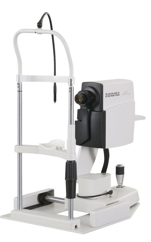
OCT retinal scan
Optical coherence tomography (OCT) is a specialised method of imaging the various layers of the retina, the optic nerve head, and the anterior portion of the eye. Similar to ultrasound, individual layers of tissue are scanned with a. safe laser, without touching the eye, and displayed a cross-section. This technology provides unique insights into the health of structures of the eye that would otherwise not be visible using standard examination techniques.
This practice believes in the highest standard of eyesore and therefore has invested in a Heidelberg engineering Spectralis OCT manufactured in Germany. This device is the preference of choice for many ophthalmologists and hospitals across the world due to it’s highly accurate and repeatable scans of the eye. The Spectralis is capable of showing 10 retinal layers and can reliably measure changes in retinal thickness as small as 1 micron.
Our OCT scan provides information about the condition of the retinal layers and optic nerve, which can identify early signs of disease. The OCT scan is particularly useful for issues concerning fluid retention and swelling in the retina, which may often occur in age-related macular degeneration and diabetes. OCT is so sensitive it can show signs of disease before you notice changes in your vision, Studies have proven that starting treatment early is the best way to save vision.
Anterior photography
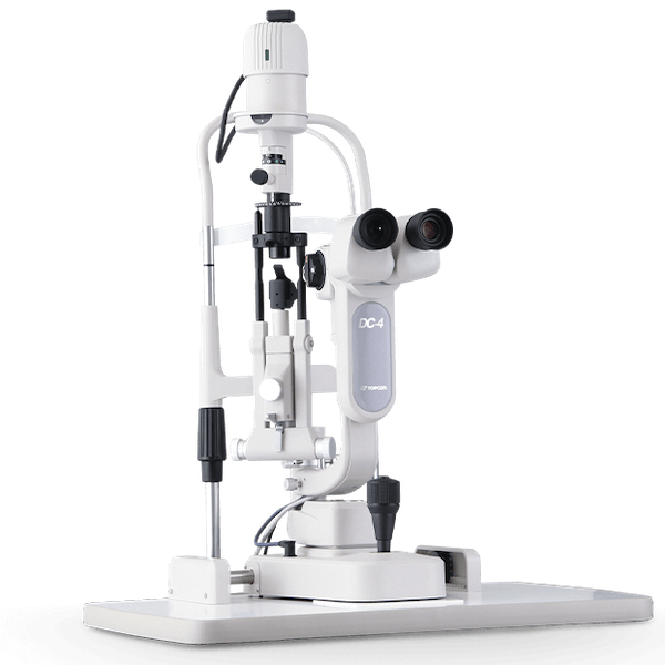
Anterior segment imaging records photos and video of vital components of the front of the eye and from deep within the eye. This information is desirable for accurate diagnosis and monitoring of pre-existing disease or newly found conditions of the eye.
This practice is equipped with the latest Topcon slit lamp with a high definition camera which captures clear and sharp still images and vivid colour video of various structures within the eye. Images in high resolution and up to 40x magnification can be obtained of the various layers of the cornea and overlying tear fluid, eyelids, lashes and deep within the eye including the lens, iris, ‘angle’ vitreous and retina. This device also uses infrared light to assess the structure and health of oil glands located within your eyelids (known as meibography) which are vital for maintaining comfortable lubrication of the eyes. These images are stored for future comparison and can be shared with other health professionals. Common eye conditions that can be imaged with high tech slit lamp include cataract, pytergium, corneal abrasion or infections, dry eye and eye lid disease, and peripheral retinal lesions.
Digital Retinal Photography
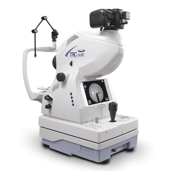
Digital retinal imaging records high definition images of the vital structures of the back of the eye. Images that can be recorded include the retina and it’s blood vessels, optic nerve tissue and the macula. Common eye conditions that benefit from digital retinal scans include age-related macular degeneration, diabetic retinopathy, epiretinal membranes, naevi (‘freckles’), glaucoma, retinal degenerative conditions, and vascular abnormalities. Other general health conditions that benefit from digital retinal scans include hypertension (high blood pressure), hyperlipidaemia (elevated cholesterol), diabetes and those taking systemic medications that may cause damage to the photoreceptors of the retina.
Visual field machine
A visual field assessment measures the extent of your peripheral vision. This test helps to diagnose and monitor certain eye conditions that effect the retina (the light sensitive layer at the back of the eye), the optic nerve (the nerve carrying information from your eye to your brain) and the integrity of the visual pathway through to the brain.
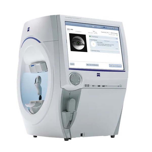
This practice can the latest ZEISS Humphrey visual field machine which is considered the ‘gold standard’ machine used by ophthalmologists worldwide. The latest software means testing time is only 3-4 minutes per eye which eliminates fatigue.
The ZEISS Humphrey visual field machine also the the ‘Esterman driving test’ program, which is the standard recommended program used for assessing visual fields in relation to maintain a driving licence.
Biometer
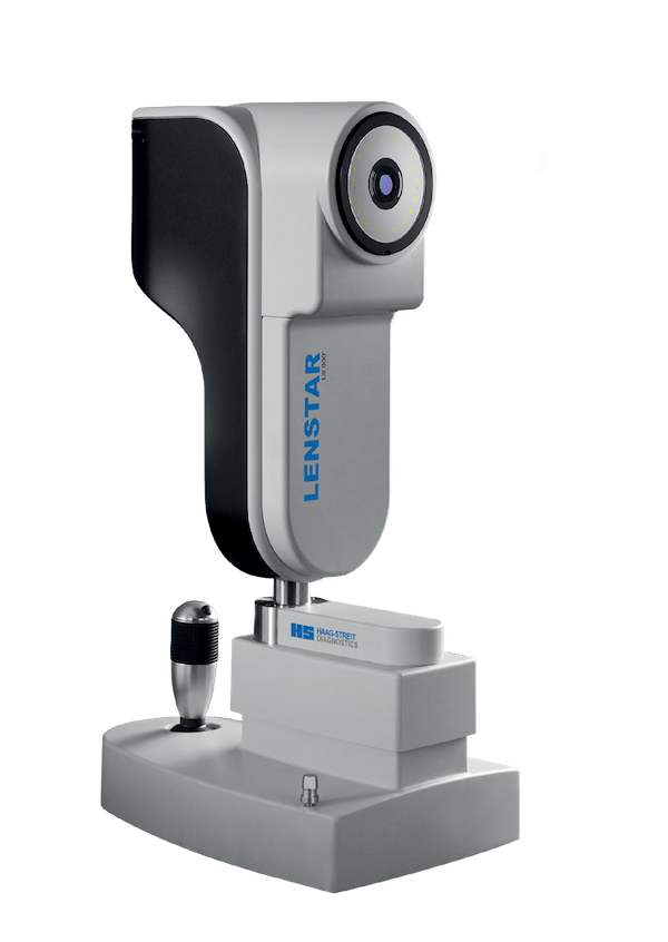
Biometry is the process of measuring the power of the cornea (shape of the front of the eye) and measuring the length of the eye. Accurate biometry measurements are critical for the appropriate management of glaucoma and progressive myopia. At this practice we use the Lenstar Myopia biometer which provides highly accurate laser optic measurements for various sections of the eye, including the thickness of your cornea and the axial length of the eye.
Using the Lenstar Myopia, we can accurately monitor the growth of the length of the eye in children with myopia, ensuring that any myopia control interventions are effective at slowing the rate of growth of the eye. We also use this device to determine if your cornea is thin compared to average corneal thickness, as thin corneas’s increase the risks of developing glaucoma when the eye pressure is higher.
Tonometer
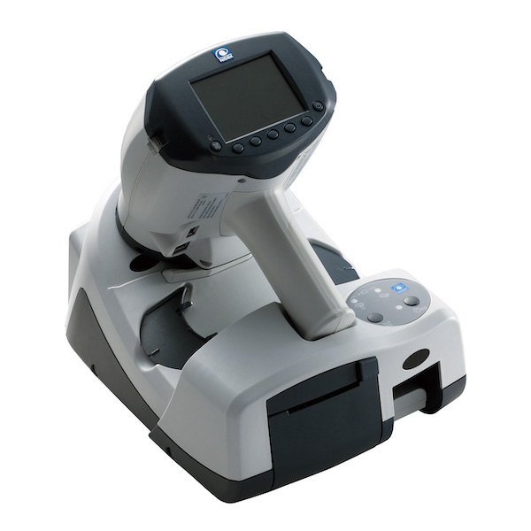
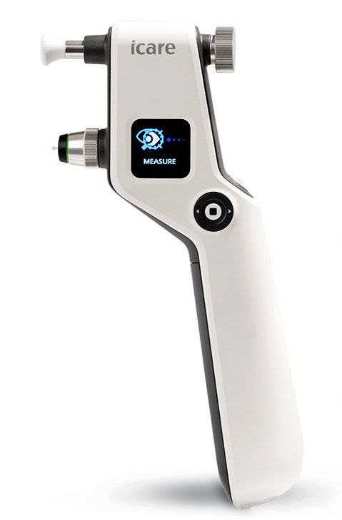
Tonometer are sensitive devices that can measure the pressure within the eye, which is particularly important for detecting or monitoring of glaucoma. At this practice we measure your eye pressure using an iCare 100 tonometer which is suitable for all patients, young and old, and provides a fast and reliable estimate of your eye pressure. This device does not require the use of anaesthetic eye drops and does not cause the sudden blinking reaction found with the ‘puff of air’ instruments of the past.
Topographer
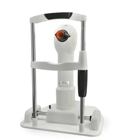
Corneal topography assesses the anterior curvature of the cornea and produces a highly precise map of the contours of your cornea. Our topographer is considered to be the ‘gold standard’ of topographers worldwide for the fitting of specialty contact lenses such as OrthoK.
The topographer in this practice can measure to an accuracy of within 2 microns, which is incredible given the width of a human hair is 75 microns. The device provides the greatest mapped area of all placid disc topographers, providing accurate measurements across the whole cornea, making it perfect for contact lens fitting, The precise mapping technology can make the fitting of custom rigid contact lenses more accurate, and can be used to monitor common progressive corneal disorders such as pytergium, and keratoconnus.
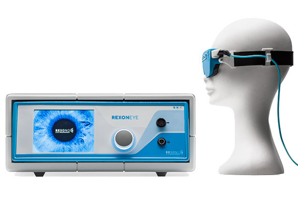
REXON dry eye machine
The Rexon-Eye is the latest dry eye innovation that uses patented Quantum Molecular Resonance (QMR) technology to treat patients with all types of dry eye disease. Rexon-Eye is the only device that can treat both aqueous deficient dry eye disease and meibomian gland dysfunction disease. The technology works by applying weal alternating electric currents at frequencies that stimulate the tear production system and increase the activity the specific glands that produce our tear fluid. This safe and pain-less procedures stimulates the metabolism and natural regeneration of the cells involved in tear production. The result is increased aqueous and tear production as well as increase quantity and quality of lipids produced by the meibomian glands. Overall, the tear film is rebalanced and the eye feels lubricated and hydrated. We are proud to be the first optometrist in Brisbane to offer this advanced dry eye treatment which was only previous only available at a few ophthalmologists.
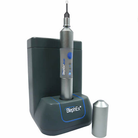
Blephex device
BlephEx micro exfoliation is a painless in office procedure that thoroughly cleanses the eye-lids, thoroughly removing excess biofilm that naturally accumulates due to the natural flora on our skin. The handpick spins a disposable micro-sponge tip that precisely cleans along the eyelid edge and lashes, removing debris and gently exfoliating the lid surface. This procedure is useful for people suffering from blepharitis, and some forms of dry disease.
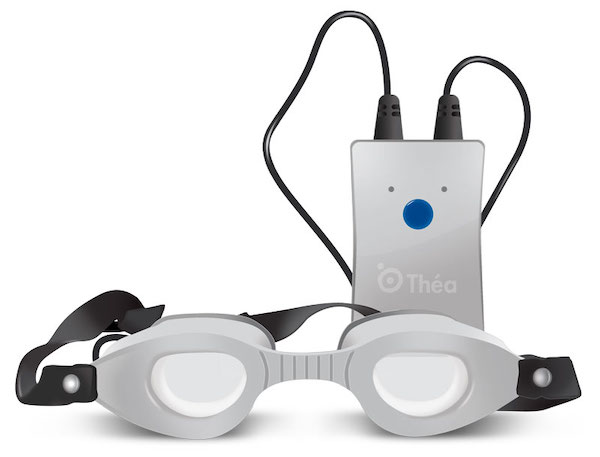
Blephasteam goggles
Blephasteam moisture chamber treatment is an in-office procedure that we perform for those with mild to moderate meibomian gland disease. The Blephasteam unit is a medical device that delivers moist heat to the eyelids in a perfectly controlled manner. The device is capable of maintaining a constant 42 degree Celsius heat for 10 minutes, which is sufficient to melt the obstructed was like oily secretions within the meibomian glads. The oily meibum secretion is the expressed with pressure applied directly to the glands relieving dry eye symptoms.
Autorefractor
An autorefractor provides an accurate estimate of your spectacle prescription just by looking into an instrument. This helps where English is not your first language or with young infants. The estimate provided can be further refined by more traditional refraction methods. This reading can be taken quickly and painlessly, however typically eyedrops will be used to control our focussing for greater accuracy.
